Characteristic Beam Patterns of X-ray Include Which of the Following
For example an 80-kVp x. Specialized X-ray sources detectors and analysis techniques have been developed to address a range of questions from the study of.

Abstract Rays Lines With Transparent Background Background Rays Burst Png And Vector With Transparent Background For Free Download Transparent Background Background Abstract Lines
Characteristics of X-ray Beam We include beam quality beam quantity and beam intensity in any discussion related to the X-ray beam.
. 1 while the x-ray absorption spectra reflect the unoccupied molecular orbitals MO. Learn vocabulary terms and more with flashcards games and other study tools. The x-ray film strip is bent into a circle of radius R.
Consists of moving horizontal and vertical lead strips 3. These two focal projections are necessarily about 90 apart in the plane normal to the filament-anode axis. Currently the most common form of beam restriction 4.
The x-ray beam is polyenergetic many energies and consists of a wide range of energies known as the x-ray emission spectrum. Energy lost is emitted as a bremsstrahlung X-ray photon. Occasionally magnification can be helpful in localising abnormalities.
Attenuation is a reduction in x-ray beam quantity intensity as a result of x-ray interactions in matter. These x-ray spectra represent local L and partial P electron density of states DOS because of the. The long axis of the femur is running perpendicular to the beam Technical factors.
Often the product of the tube current and exposure time is considered as one entity the mAs milliampere-second. The useful beam emerges from the port window. Start studying Chapter 8 Dental X-Ray Characteristics.
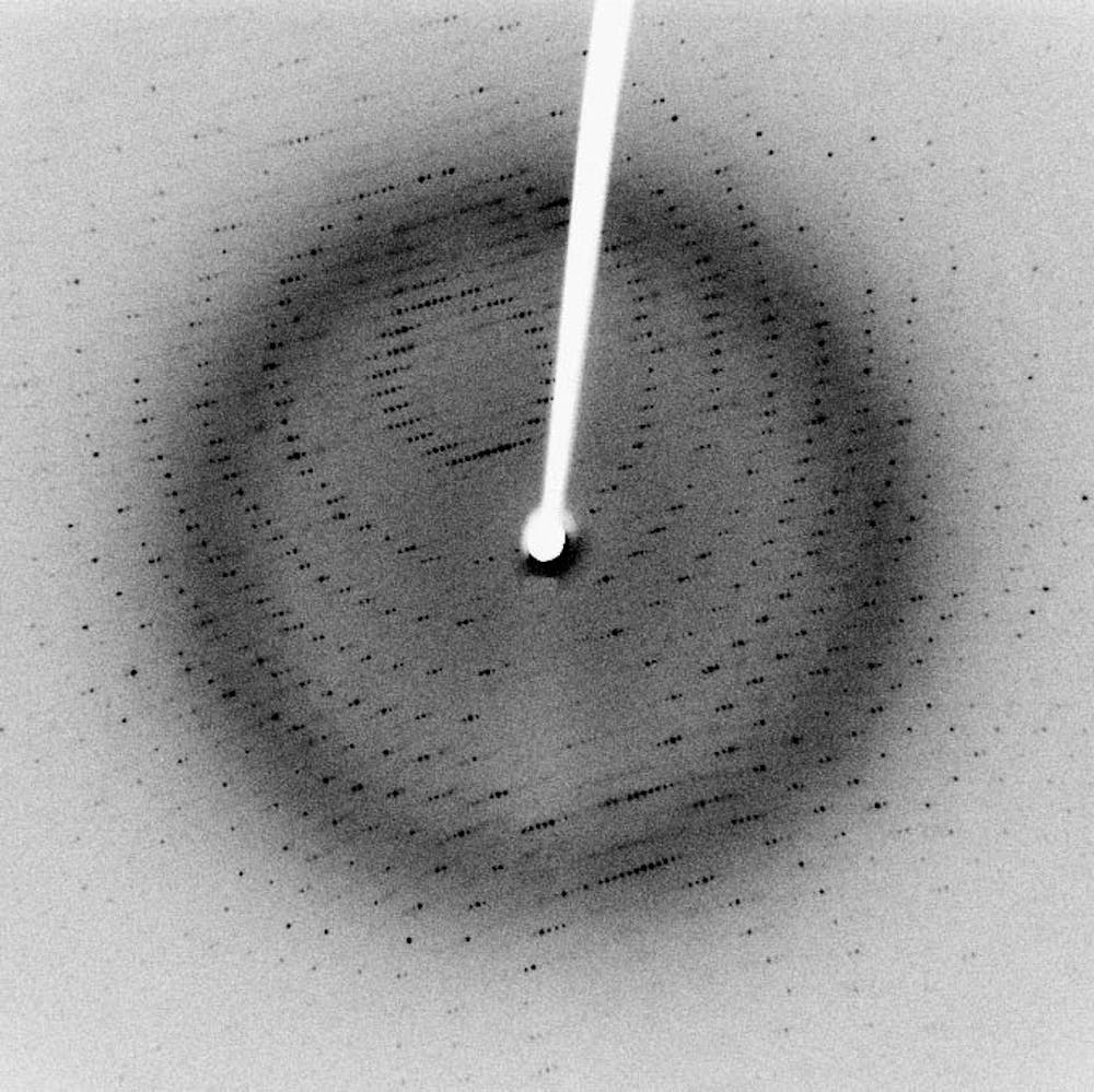
The pattern-transferring processes include four key steps. Braggs law is used to determine a crystal parameters from its characteristic x-ray pattern. This is the result of x-ray photons being absorbed or scatter out of the x-ray beam.
X-ray emission spectra of solids and molecules are methods of measuring electronic structure of matter 15. Approximately 80 of the population of X-rays within the X-ray beam consists of X-rays generated in this way. X-rays travel in straight lines.
2Heating causes thermionic emission to occur- boiling off of electrons specifically outer shell electrons. X-Ray beams that are parallel with wide projection of the filament have a focal shape of a line. The kV mA and exposure time are the three major selectable parameters on the x-ray generator control panel that determine the x-ray beam characteristics.
A set of silicon mirrors coated with silicon carbide with an incidence angle of 132 mrad are located in the front-end enclosure of the Near Experimental Hall NEH which feeds the hard X-ray beam to all LCLS hard X-ray hutches. The sketch should show. The x-ray emission spectra reflect the occupied electronic structure as shown in Fig.
From the focal spot the xray diverge into space forming the cone-shaped primary xray beam. X-Ray beams that are parallel with the narrow projection of the filament have an approximate focal shape of a square which is usually labeled as a spot. Which of the following is are characteristics of the x-ray tube.
Search for Bragg peaks by using not a monochromatic x-ray beam but one containing wavelength for up to λ1. Body parts further away from the detector are magnified compared with those that are closer. Coherent X-ray beam metrology enabled by a.
Three characteristics of the x-ray beam. X-rays travel in straight lines and a beam of X-rays diverges from its source. When a free electron fills the shell a x-ray photon with energy characteristic of the target material is emitted.
May be manufactured with a positive beam limiting device. Designs of the x-ray facility which tries to take into account of the synchrotron beam characteristics and of the ease in operation as well as provisions for combining the x-ray instrumentation. X-ray energy is measured in kiloelectron-volts keV 1000 electron volts.
Single-shot and acquired with a LiF crystal imaging detector. Electron path in x-ray tube x-ray tube target x-rays going from target into powder crystal camera slits that define x-ray beam going to sample under study sample beam catcher for directly transmitted beam diffracted x-ray beam film strip. Bremsstrahlung Braking radiation.
Refers to the number of x-rays produced in the dental x-ray beam. Consider the following types beam restrictors and match them to their respective characteristics 1. Combination of the number of x-ray photons quantity and the energy of each photon quality.
Used to describe the energy or penetrating ability of the x-ray beam. These different techniques are generally termed photolithography X-ray lithography electron beam lithography or ion beam lithography depending on the radiation employed. Imaginary perpendicular ray at its center is called central ray.
Which of the following photon interactions contribute to the attenuation of. When an outer shell electron moves to fill the space created in the inner shell energy in the form of an x-ray photon is emitted. The following methods are used to relax the constrains in order to achieve diffraction peaks 1.
Instrument overview The XCS instrument operates in the hard X-ray regime above 4 keV. Characteristic x-rays are caused by the ejection of an electron from an inner shell of an atom hit by the incident x-ray. When an electron passes near the nucleus it is slowed and its path is deflected.
The defining characteristics of X-raystheir ability to penetrate optically opaque materials their wavelengths of atomic dimension the high energy of individual X-ray photonslead to a wide range of industrial medical and scientific applications. The cathode assembly receives both low and high voltages. The lowest energies are always approximately 15 to 20 keV and the highest energies are always equal to the kVp set on the control panel.
The target material should have a high atomic number and a high melting point. 1 Current will heat the tungsten filament. 3 Electrons that are emitted form a space charge- cloud around the filament.
Centre to the knee joint 15-20 cm distal to the apex of the patella or at the tibial tuberosity if the patella is affected by certain injury patterns. The intensity patterns produced were obtained in situ with a. Classified as the simplest form of a beam restrictor 2.
The cross section of the xray beam at the point where it is utilized is called the radiation field. Laue method Fix the orientation of the single crystal. Superior to include the distal femur.
Dissemble the powder crystal camera if it is not already done enough. Cross section of xray beam is called the radiation field. 1 solution-based wet chemical etching procedures 2 dry etching in a reactive plasma 3 doping using ion implantation techniques.
Therefore it is essential to distinguish between these concepts and understand the factors that influence them. Common targets used in x-ray tubes include Cu and Mo which emit 8 keV and 14 keV x-rays with corresponding wavelengths of 154 Å and 08 Å respectively.
Abdomen Xray Signs

Chest Radiograph Radiology Reference Article Radiopaedia Org

Operator Error The Moire Pattern Seen In This Knee Image Was Caused By Download Scientific Diagram

Initial Chest X Ray Showing Reticulonodular Pattern With Midzone Download Scientific Diagram

X Ray Diffraction Pattern Of A 316l Stainless Steel And B As Welded Download Scientific Diagram

Initial Chest X Ray Showing Reticulonodular Pattern With Midzone Download Scientific Diagram
![]()
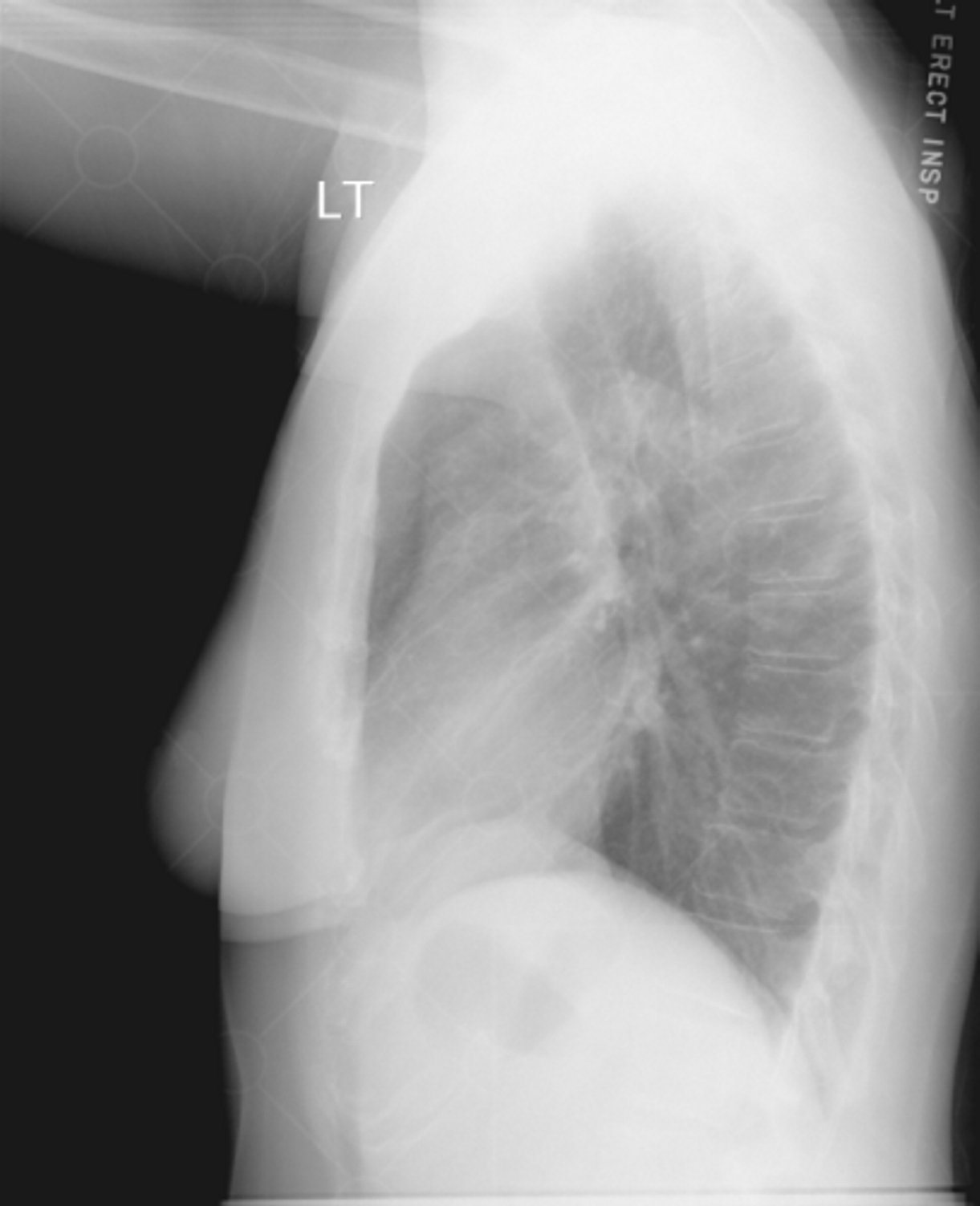
Normal Chest X Ray Anatomy Tutorial Kenhub
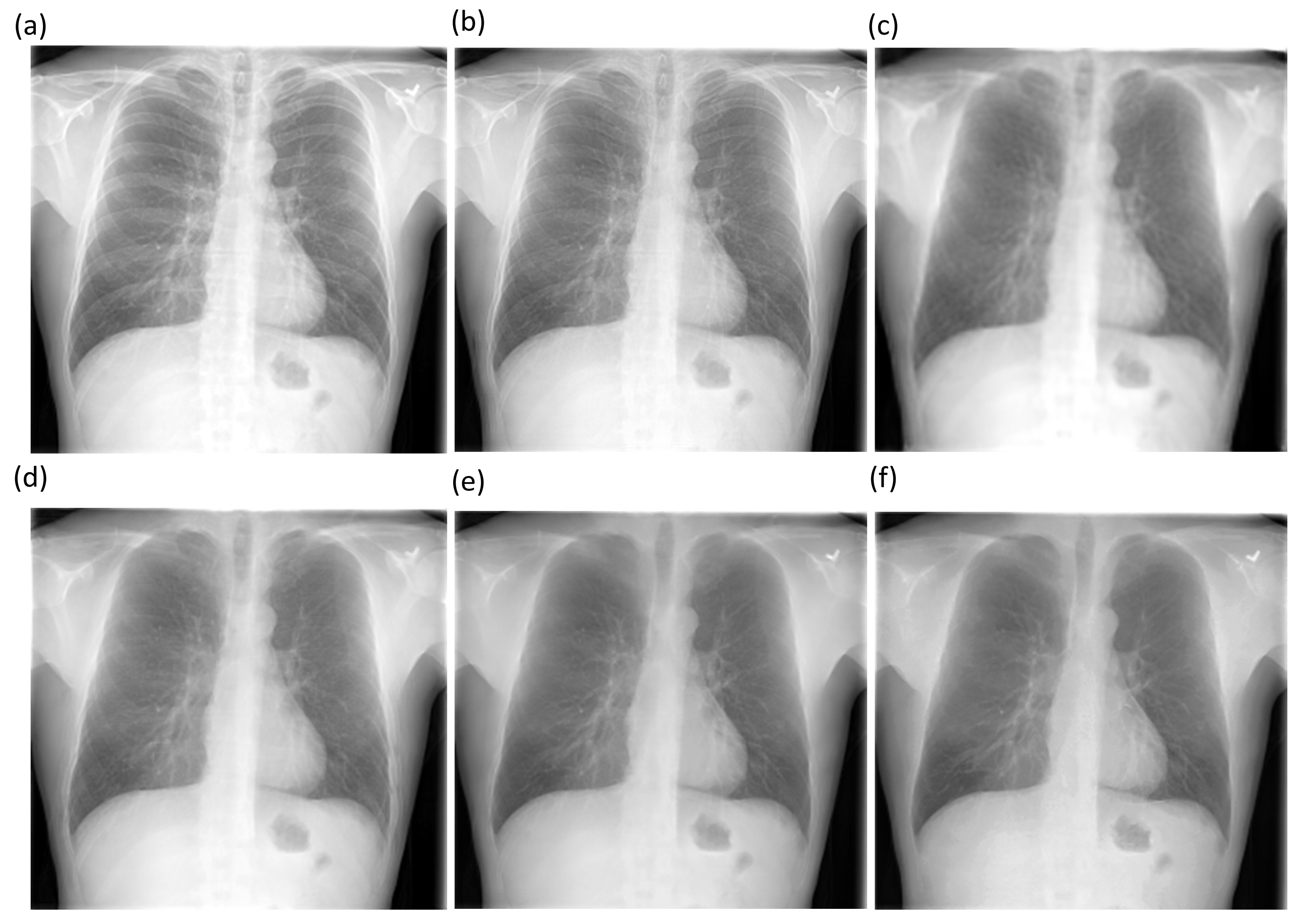
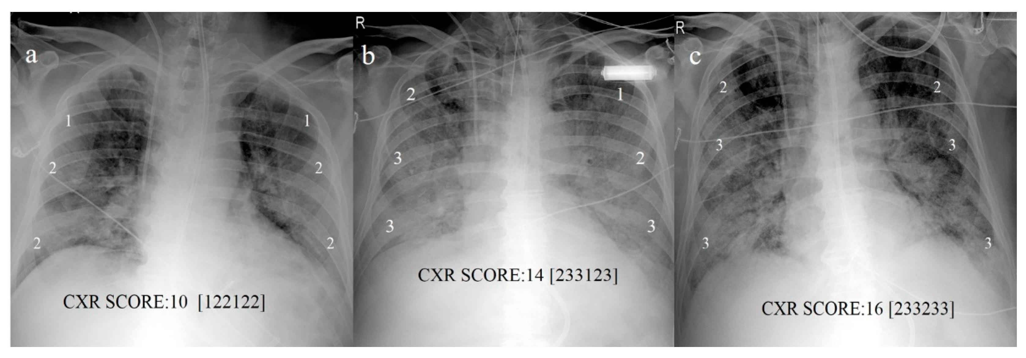
Diagnostics Free Full Text Chest X Ray Bone Suppression For Improving Classification Of Tuberculosis Consistent Findings Html

The Left Lower Lobe Is Reduced In Volume And Opaque On Lateral Projection The Oblique Fissure Is Di Radiology Medical Radiography Human Anatomy And Physiology

X Ray Diffraction Patterns Of Manganese Oxide Samples Download Scientific Diagram
Digital Radiography Image Artifacts Radiology Suny Upstate Medical University
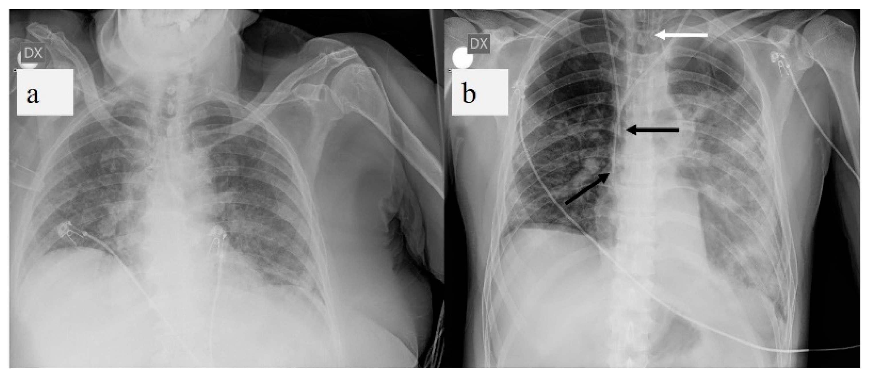
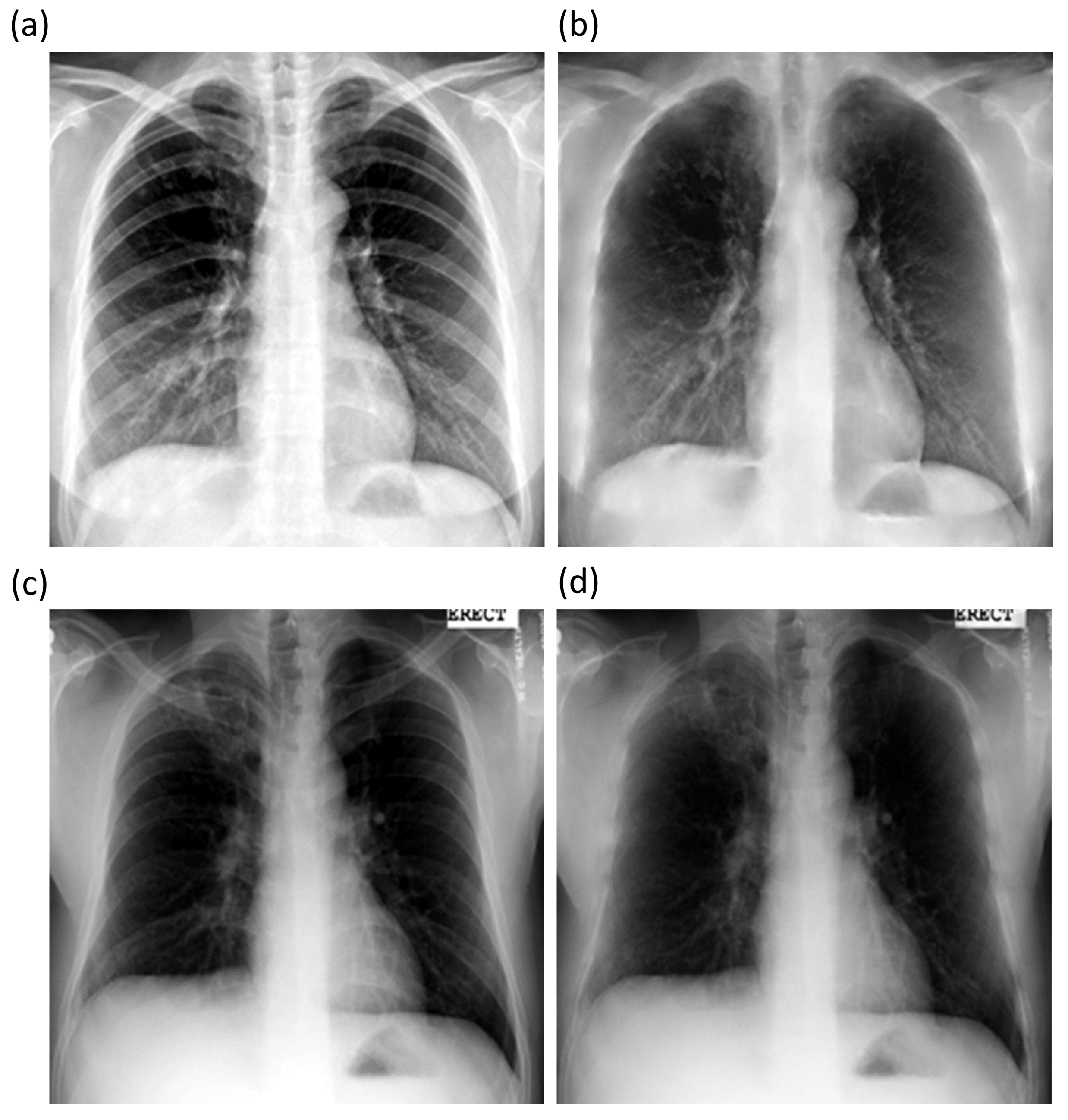
Diagnostics Free Full Text A Pictorial Review Of The Role Of Imaging In The Detection Management Histopathological Correlations And Complications Of Covid 19 Pneumonia Html
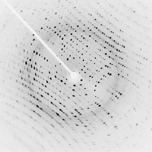
X Ray Diffraction University Physics Volume 3
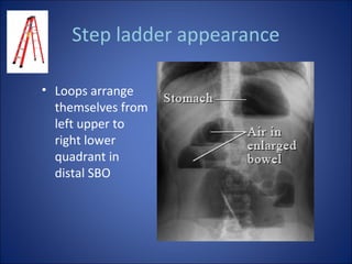
Abdomen Xray Signs
Diagnostics Free Full Text Chest X Ray Bone Suppression For Improving Classification Of Tuberculosis Consistent Findings Html
Diagnostics Free Full Text A Pictorial Review Of The Role Of Imaging In The Detection Management Histopathological Correlations And Complications Of Covid 19 Pneumonia Html

The X Ray Diffraction Pattern Of The Zinc Oxide Nanoparticles Prepared Download Scientific Diagram

Elevated Right Hemidiaphragm Hepatomegaly Human Body Anatomy Radiology Body Anatomy
Explainer What Is X Ray Crystallography


Comments
Post a Comment